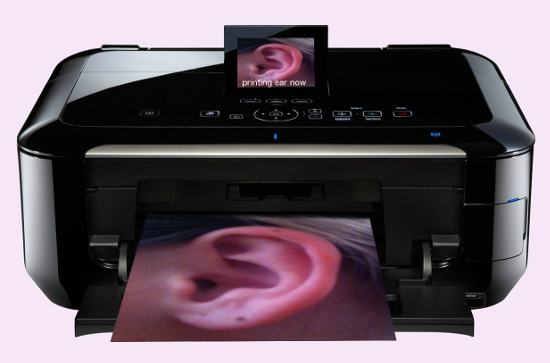
Ten years ago, I posted an article about human cell research that would one day give Plastic Surgeons the ability to print the cartilage that gives ears and noses their shape. Well that day is closer than ever. Scientists at Swansea University in Wales have announced a new research project to determine the ideal combination of cells needed to grow new cartilage. Today’s San Francisco Plastic Surgery Post reveals the latest progress made toward custom printing body parts.

Reconstructing the ear for congenital absence of the ear or after severe trauma may have just gotten a little bit easier.
3D Printing
3D printers have been around for a while, but the ability to 3D print body parts is relatively new. The prospect of transforming abnormal anatomy into functional form has long been the dream of plastic surgeons. The advances in technology and science might provide the necessary tools to “print” the building blocks for replacement cartilage.
Where is Cartilage Found?
Cartilage is found in the ears, nose, ribs and joints. Unfortunately, the availability of cartilage for reconstruction is very limited. Common donor sites are the septum of the nose for firm straight struts, the concha of the ear for soft curved pieces and the ribs for bulkier blocks. The amounts of available cartilage are limited, and the donor sites are subject to compilations. The ability to print cartilage for structural reconstruction of the ear, nose and possibly joints would be a game changer.
Correcting Microtia
The need for 3D printed cartilage is most obvious for microtia or anotia, the congenital absence of the ear. Unlike Otoplasty for Prominent Ears, where the problem is the shape of the cartilage, with microtia there is a lack of cartilage. The current treatment for reconstruction is to remove several blocks of cartilage from the junction between the ribs and sternum, stack them together and then carve the desired shape. The process of harvesting the cartilage, carving the needed shape and covering it with skin is labor intensive and time consuming. It’s far from ideal, but it’s the current gold standard.
3D Printing An Ear

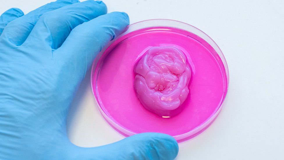
Human cells may soon be used to 3D print ear cartilage. Hopefully, with a better shape than that shown in the photo used in the BBC news article.
Once the optimal combination of cells is determined, a 3D printer could be used to construct the required supportive scaffolding. For ear reconstruction, a digital scan of the opposite ear could be used to construct the perfect mirror image. Printing would be performed in a laboratory outside the operating room, which would significantly reduce the operating time as well as donor site complications.
Prominent Ears
Ears that stick out are much easier to fix than no ears. Last week’s Otoplasty Post gives several examples. If you are interested in Otoplasty call (925) 943-6353 and schedule a private ear shaping consultation.
Previous Post Next Post